
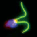
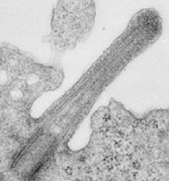
 Primary cilia are protrusive microtubule based-organelles that resemble flagella of protists. Almost every cell in the human body is equipped with a solitary primary cilium. First image on right shows electron microscopic image of a mouse primary cilium (Liem et al., 2012). The second is an immunofluorescence image of the green alga Chlamydomonas with flagella marked in green (generated by students of the EMBO Practical Course ‘The Molecular Genetics of Chlamydomonas’).
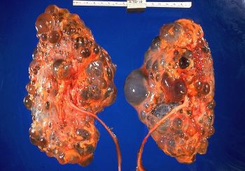 Cilia have been mostly ignored by mainstream biomedical science for decades – they were considered to be vestigial organelles lacking important functions in mammals. They have recently returned into the spotlight when it was discovered that many human diseases, so called ciliopathies, are caused by dysfunction of primary cilia. Image on left shows cystic kidneys – a common symptom of many ciliopathies.
 Interestingly, many components of Hh signaling are present in cilia. Ptch is concentrated in the ciliary membrane when the pathway is inactive, whereas Smo, Gli, and SuFu move into the cilia when the pathway is turned on (the image on left shows Gli2 localized at tips of cilia – marked with acetylated tubulin – in cells stimulated with a Hh agonist). Although some progress has been made in identifying sequences that target membrane proteins to cilia, the mechanism of the transport of soluble proteins into these organelles remains mysterious.
Questions that we are trying to resolve:
How are Gli proteins targeted to cilia and what are the proteins responsible for their transport to cilia tips?
More generally – why do some soluble proteins remain concentrated in cilia, while others are excluded from the ciliary compartment?
What is the function of ciliary transport of Gli proteins?